Distinguishing the malleus from the incus or tracing the spiral of the cochlea is difficult enough from a flat diagram. A physical 3D replica turns abstract anatomy into a tactile, spatial experience you can hold, rotate, and disassemble. Whether you are training to become an audiologist, preparing patient education materials, or studying for a medical exam, the right model makes the difference between memorizing labels and truly understanding form.
I’m Min — the co-founder and writer behind Gadgets Feed. I have spent hundreds of hours researching anatomical teaching aids, comparing magnification scales, material durability, and part-count dissectibility to separate the truly educational models from the purely decorative ones.
This guide breaks down the best models for every use case and budget. My goal is to help you confidently select a best anatomical ear model that fits your learning or professional needs without wasting time on poorly constructed replicas.
How To Choose The Best Anatomical Ear Model
The wrong ear model either oversimplifies the tiny bones into a lump of plastic or presents a fragile replica that breaks on first assembly. Three core factors separate a useful teaching tool from a dust-collecting paperweight.
Magnification Scale and Part Count
Life-size ear models are compact but force you to squint at the stapes and incus. A 3X enlargement is a solid starting point for most students. A 5X or 8X scale reveals the cochlea’s internal spiral and the semicircular canals with far greater clarity. Models that break into at least three parts — external, middle (with removable ossicles), and inner ear — offer a much richer learning experience than solid single-block replicas.
Materials and Build Quality
Medical-grade PVC is the standard for durable, hand-painted anatomy models. It resists chipping and cleans easily with a damp cloth. Silicone models serve a different purpose: they are soft enough to pierce with earring posts, making them ideal for jewelry display or piercing practice but useless for studying fine anatomical detail. Laser-etched crystal glass models are decorative conversation pieces, not serious study tools.
Intended Use Case
An audiology student preparing for exams needs a highly detailed, multi-part model with a numbered key. A physical therapist explaining Eustachian tube function to a patient needs a model that is large enough to point at but does not require disassembly. A jewelry maker displaying handmade earrings needs a simple, soft ear form that will not scratch metal posts. Match the complexity of the model to the depth of understanding you require.
Quick Comparison
On smaller screens, swipe sideways to see the full table.
| Model | Category | Best For | Key Spec | Amazon |
|---|---|---|---|---|
| Axis Scientific 5X Enlarged | Premium | Audiology study & classroom | 5X life size, 3 parts | Amazon |
| EVOTECH 4-Part 3X Enlarged | Premium | Medical student dissection | 3X size, 4 parts | Amazon |
| GPI Anatomicals Full-Size | Premium | Doctor-patient education | Life size, labeled base | Amazon |
| VEVOR 5X Enlarged | Mid-Range | Detailed classroom display | 5X size, 3 removable parts | Amazon |
| EVOTECH Inner Ear Labyrinth | Mid-Range | Inner ear specialization | 8X size, 2 parts | Amazon |
| Cindeer Silicone Ear Set | Budget | Jewelry display & piercing | Soft silicone, 4-pack | Amazon |
| XINDAM Crystal Glass Ear | Budget | Decorative gift | Laser-etched glass cube | Amazon |
In‑Depth Reviews
1. Axis Scientific Human Ear Model, 5 Times Enlarged 4 Dimensional Model with 3 Parts
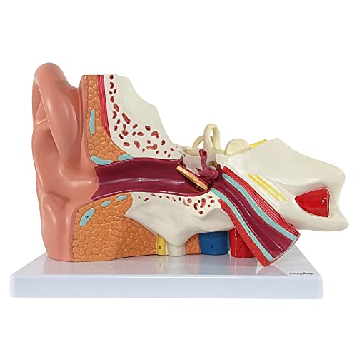
The Axis Scientific model strikes the best balance between magnification and structural detail. At 5X life size, the external ear, middle ear cavity, and inner ear labyrinth are large enough to inspect without a magnifying glass. The model dissects into three pieces, allowing you to separate the major regions and study the relationship between the tympanic membrane and the ossicles.
Every structure is hand-numbered, and the included full-color manual uses actual photographs of the model rather than generic diagrams. This means you can directly match the number on the plastic part to the image in the guide. The dense PVC plastic feels substantial and withstands repeated assembly and disassembly in a classroom environment.
Audiology students and professors consistently praise its clarity and accuracy. The base is wide and stable, keeping the model upright during demonstrations. If you need one model to serve both personal study and patient or student education, this is the most complete package available.
Why it’s great
- Hand-numbered parts match a photo-based manual for easy identification
- Sturdy PVC construction with a stable base that resists tipping
- Three-part dissectibility reveals inner, middle, and external ear clearly
Good to know
- Premium price point may exceed a casual student’s budget
- Individual ossicles are not separable from one another
2. EVOTECH SCIENTIFIC Human Ear Model Anatomy, 4 Parts 3X Enlarged
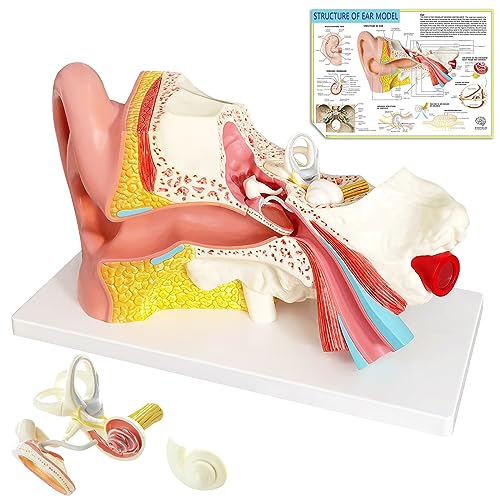
The EVOTECH 4-part model is unusual in its class because it splits into more pieces than the typical 3-part design. The tympanic membrane and the labyrinth can both be removed, giving you access to the intact cochlea and the bone labyrinth separately. At 3X life size, the structures are still comfortably visible for group demonstrations.
The hand-painted PVC finish is detailed enough for a college anatomy and physiology class. The base holds the model securely, and the overall weight of just over 3 pounds gives it a reassuringly solid feel. Medical students in clinical training have reported that the model helps bridge the gap between textbook diagrams and real surgical anatomy.
The main limitation is that all three ossicles are fused into a single piece. A student who needs to manipulate the malleus, incus, and stapes independently may find this frustrating. However, for the price, the sheer number of detachable components makes this the strongest value proposition in the mid-range segment.
Why it’s great
- Four separate parts provide deeper dissectibility than most competitors
- Removable labyrinth and tympanic membrane for focused study of inner ear
- Solid PVC construction with accurate hand-painted details
Good to know
- Ossicles are fused into one unit, not individually removable
- Some users reported small assembly pegs can be fragile
3. GPI Anatomicals – Ear Model, Replica of a Normal Ear
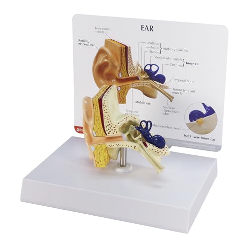
The GPI Anatomicals model is a life-size replica designed specifically for patient education in a clinical setting. It does not attempt to be an enlarged dissection model. Instead, it shows the normal human ear at actual scale with the semicircular canals, cochlea, auditory ossicles, tympanic membrane, and the tensor tympani muscle clearly painted and labeled.
The included information card and display base make it suitable for a doctor’s office desk or an exam room shelf. Physicians and audiologists have noted that it is particularly effective for demonstrating digital otoscopy technique because the proportions match a real patient’s ear. The model measures just over 3 inches tall, so it does not dominate a desk.
This is not a model for students who need to disassemble and reassemble the ear repeatedly. The individual pieces do not come out. But for explaining anatomy to a patient who has never seen their own eustachian tube, the accurate scale and clear labeling are far more effective than a poster or a diagram.
Why it’s great
- True-to-life scale ideal for otoscopy demonstrations and patient education
- Includes a labeled information card and a sturdy display base
- American-designed and manufactured with over 40 years of anatomical model expertise
Good to know
- Non-dissectible — no removable parts for hands-on study
- Small size may be difficult for group classroom viewing at a distance
4. VEVOR Human Ear Anatomy Model, 5 Times Enlarged
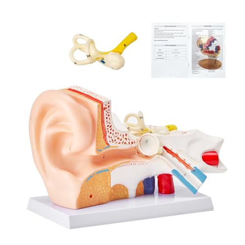
The VEVOR model is a 5X enlarged replica that emphasizes visual clarity through color coding. Each anatomical region — external, middle, and inner ear — is painted in a different color, making it immediately obvious where one structure ends and another begins. The numbers and letters printed on the surface correspond to a labeled key for self-guided study.
Two major structures are removable, letting you lift off parts of the middle and inner ear to see the layered arrangement beneath. The ABS base provides excellent stability for a model that measures nearly 12 inches across. The high-quality PVC has minimal odor compared to cheaper plastic alternatives, which is a real advantage in an enclosed classroom or lab.
Some customers have reported missing the tympanic membrane and ossicles in their shipment, which points to occasional quality control gaps. When the model is complete, it offers outstanding detail for its price tier. If you receive a fully intact unit, the color-coded approach makes it one of the easiest models to teach from.
Why it’s great
- Color-coded anatomy simplifies identification of outer, middle, and inner ear regions
- Dismountable design reveals overlayed structural layers
- Low-odor PVC material is suitable for indoor classroom use
Good to know
- Some units have arrived missing small parts like ossicles or the tympanic membrane
- Middle ear detail has been described as slightly simplified by some reviews
5. EVOTECH SCIENTIFIC Inner Ear Model Labyrinth Model 2 Part 8X
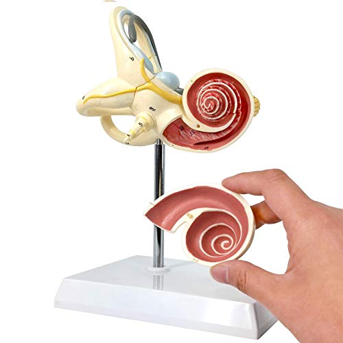
This model from EVOTECH is hyper-specialized: it focuses entirely on the inner ear labyrinth at an 8X enlargement. The bone labyrinth and membrane labyrinth are both represented, and the cochlear cover can be lifted to reveal the internal spiral structure. The semicircular canals and vestibule are also open, displaying the saccule and utricle.
Hand-painted PVC and a compact base make this easy to place on a desk or carry to a study group. Medical professionals in clinical settings have commented that the 3D representation helps patients grasp inner ear disorders like benign paroxysmal positional vertigo far more effectively than any drawing. The two-part design is simple enough to reassemble quickly during a consultation.
The trade-off is that this model shows only the inner ear. If you need to study the external ear, tympanic membrane, or middle ear ossicles, you will need a separate model. The fragile plastic pegs that hold the detachable cover in place require careful handling to avoid breaking.
Why it’s great
- 8X enlargement provides an exceptionally clear view of the cochlea and semicircular canals
- Opened cochlear cover reveals internal spiral structure for deep study
- Excellent tool for explaining vertigo and balance disorders to patients
Good to know
- Only covers the inner ear — does not include external or middle ear structures
- Detachable parts use small plastic pegs that can break under rough handling
6. Cindeer 4 Pcs Silicone Ear Model with Acrylic Stand

This set of four soft silicone ears from Cindeer has a completely different purpose from the PVC anatomy models above. Each ear is made from pliable white rubber that accepts an earring post without resistance and self-heals almost completely when the post is removed. This makes them perfect for jewelry display or for practicing piercings before working on a real ear.
The included acrylic stand holds each ear upright, so studs and drop earrings are visible from all angles. Jewelry makers have found that displaying bead earrings on these forms sells better than laying them flat on a table. The silicone material also mimics the feel of real cartilage well enough for basic piercing technique practice.
These are not anatomical study aids. There are no labeled structures, no dissectible parts, and no representation of the inner or middle ear. But for their intended use — a cheap, reusable, and soft ear model for display or practice — they outperform any PVC model.
Why it’s great
- Soft silicone accepts earring posts without damage and closes up after removal
- Four ears with an acrylic stand provide plenty of display or practice space
- Extremely affordable for jewelry makers and piercing students
Good to know
- No anatomical detail — completely unsuitable for medical study
- White silicone can show visible puncture marks after many uses
7. XINDAM 3D Human Ear Anatomical Model in Crystal Glass Cube
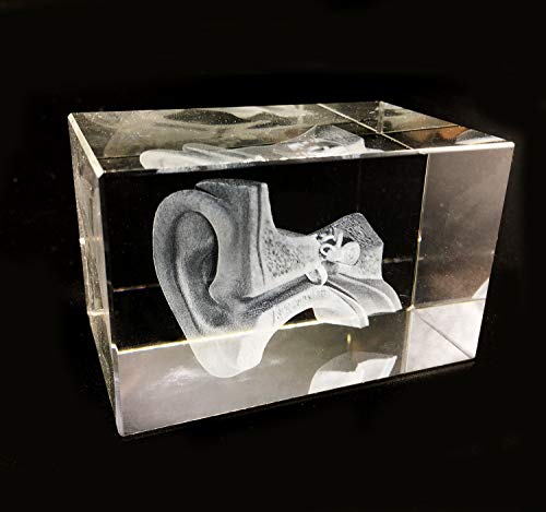
The XINDAM model is a laser-etched glass cube measuring roughly 3 inches on each side. A laser has burned a three-dimensional representation of the human ear’s internal anatomy into the interior of the crystal, creating a floating ghost image that catches light and rotates as you turn the cube. It comes without an LED base, though one can be purchased separately online.
As a teaching tool, its utility is limited. The etched lines cannot convey the texture, color coding, or dissectible layers of a PVC model. However, as a desk paperweight or a gift for a medical student or anatomy enthusiast, it is visually distinctive. The glass is solid and heavy at 1.25 pounds.
If you are looking for a serious study aid, look elsewhere. If you want a conversation piece that shows appreciation for the field of otology, this cube is a thoughtful and unusual choice. It fills a niche that no other model on this list occupies.
Why it’s great
- Unique laser-etched glass construction is visually striking and durable
- Works well as a desk ornament or gift for anatomy enthusiasts
- Compact 3-inch size fits easily on any shelf or desk
Good to know
- Not a functional teaching model — no dissectible parts or labeled structures
- LED base not included and must be purchased separately
FAQ
What magnification scale is best for studying the ossicles?
Can an anatomical ear model be used for medical exam preparation?
Why would I choose a life-size model over an enlarged one?
Final Thoughts: The Verdict
For most users, the best anatomical ear model winner is the Axis Scientific 5X Enlarged 3-Part Model because it combines the highest magnification usable for general study with a dissectible design and a photo-based manual. If you need a deeper focus on the inner ear labyrinth, grab the EVOTECH 8X Inner Ear Model. And for a life-size clinic display that aids patient communication, nothing beats the GPI Anatomicals Full-Size Model.